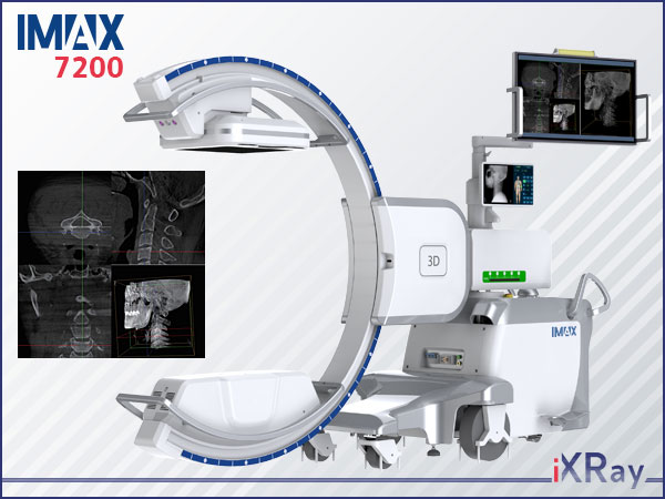
IMAX 7200
Lead the Trend of 3D-imaging
Futures & Benefits
Maximizing surgical confidence with 3D imaging
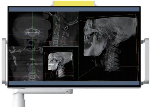
● Wide medical monitor showing images with high brightness and high contrast.
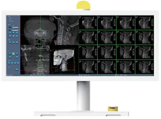
● Integrated monitor presents real-time images for easy image reading, relieving the burden of memory.
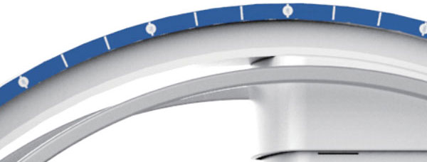
● Fully motorized movements control over your procedures.
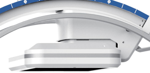
● Flat-panel detector with excellent imaging.
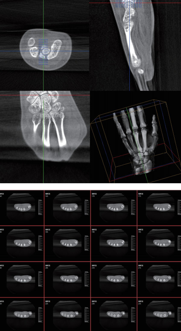
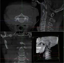
● lntraoperative 3D imaging and CT-like sectional data for more confidence in OR.
lntraoperative 3D confirms your results
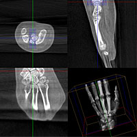
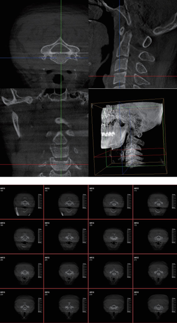
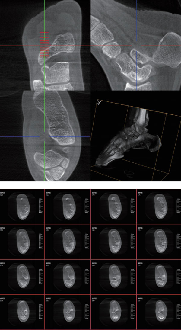
Intraoperative 3D imaging and CT-like imaging provides precise information from every angle during the surgical procedures — pinpoint anatomical structures, implants and screws more confidently.
Large volume of 3D imaging
Delivers a 3D image covering a volume of a vertical cylinder more information would be seen in one volume:
● Seven cervical vertebrae
● Seven thoracic vertebrae
● Five lumbar vertebra
● Bilateral iliosacral joints
● Femur head and unilateral pelvis
Easy 3D setup
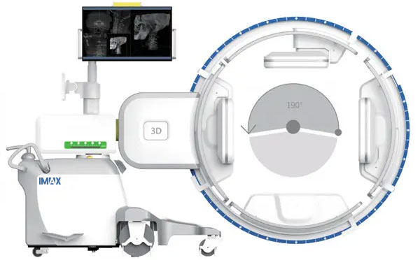
Thanks to wide space of C-arm, 3D scan procedure takes around 60s for complete information, which translates into reduced surgery time for clinical work.
More efficient workflow
Intuitive intraoperative 3D evaluation avoids unnecessary postoperative CT scans and corrective surgery, saving time and costs.

Complete 3D scan
With iso-centric scan technology, orbital movement in a motorized 3D scan from any direction giving you complete, highly accurate 3D information in outstanding quality.
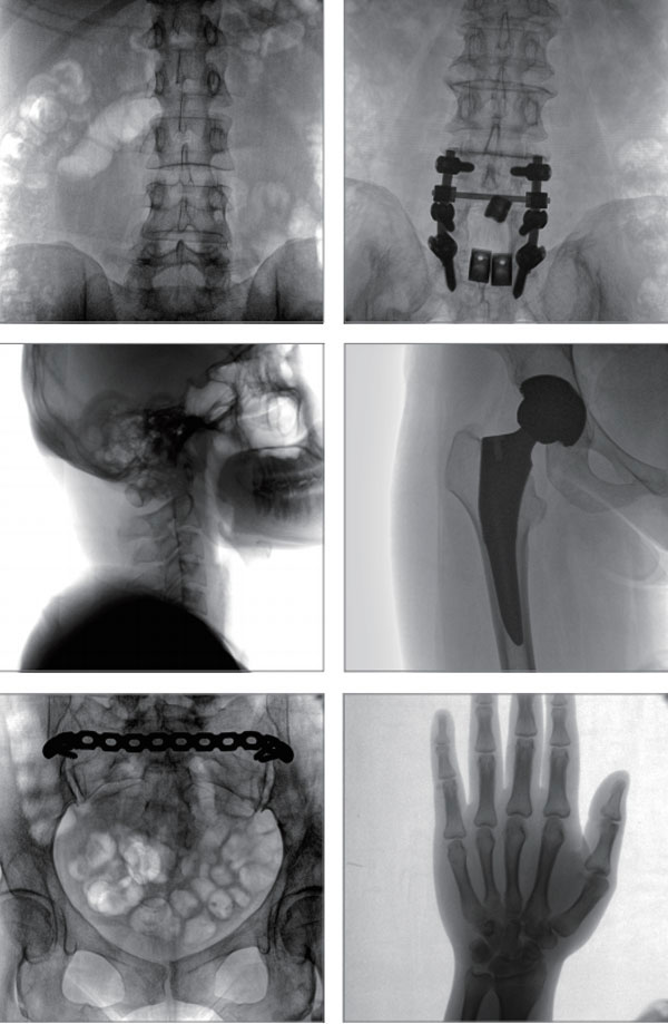
Excellent 2D, 3D image quality
Smart dose control
Adjust dose dynamically during fluoroscopy with intelligent hardware and closed-loop softw-are algorithms and minimize dose while optimizing image quality.

High frequency X-ray generators
Self-developed high frequency x-ray generator achieves outstanding images by adjustable pulse frequency, even in obese patients and dense tissue.
Get connected to navigation
Seamless navigation port connects the3D C-arm to the navigation or robotic guidance system wirele-ssly.Image-guided surgery allows for less invasive approaches and more decision-making confidence within the OR.

Flat panel detector technology
Large amorphous silicon flat detector offers large, crystal-clear 2D image and high-resolution 3D reconstruction.Every tiny anatomical structures and implants become visible.
Exceptional ease of use
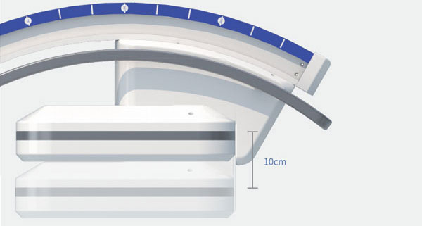
Vertical movement of flat panel detector
Adjustable movement of flat panel detector allows for intraoperative fine-tuning of the distance between the detector and the subject, to receive more x-ray signal for clearer imaging based a certain dose.

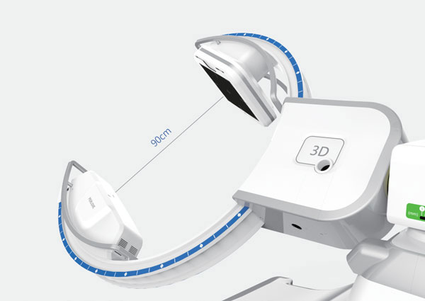
Wide space C-arm
With a tube-detector distance, Skybow offers plenty of room for easy setup and quick positioning.
Multiple infection control
No external cable between C-arm and chassis makes it easy to be covered with sterile drapes.The surface of C-arm features anti-microbial paint, maintaining a high level of infection control.
All motorized movement control
All motorized movements in 5directions move theC-arm into the exact position desired safely and stably.
Wireless footswitch
Optional footswitch creates greater freedom in OR, and avoids damage caused by trample to cable.
Multiple screen display
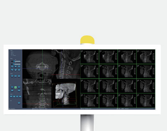
Medical color monitor
Wide medical color monitor with high resolution, brightness and contrast provides ultimate reading experience.
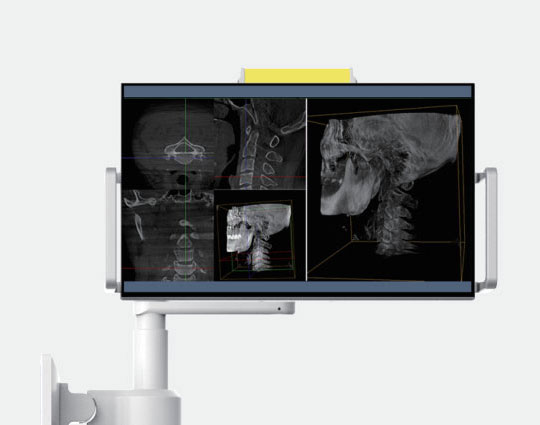
lntegrated review monitor
lntegrated medical color monitor on the C-arm body offers near-table reviews,relieving memory burden.
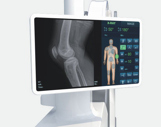
lntuitive touchscreen interface
lntuitive touchscreen control center creates quick control and sterile environment, saving time during procedures.
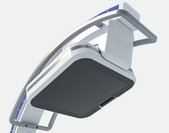
Removable grid
Remove grid by barehand to reduce dose in pediatric and other dose-sensitive procedures.
Steps to create a 3D-image
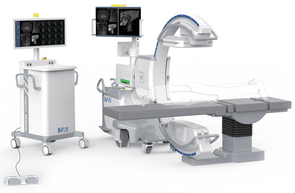
01 A right operation table
02 Patient positioning
03 Use exposure protection
04 Set up the C-arm
05 Positioning in the isocenter with laser
06 Parameter setting
07 Collision check
08 Start the scan with footswitch

Low dose mode
Reduce dose exposure significantly for particularly dose-sensitive procedures, e.g. in pediatrics.
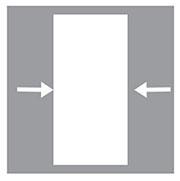
Collimators preview
Preview and positioning collimators in exposure-free conditions.

Realtime dose monitoring
Intuitive dose display for easy assessments of patient dose (DAP) and effective control.
3D-images
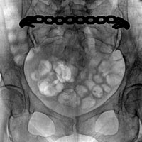
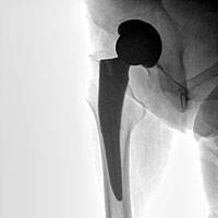
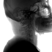
2D-images
