IMAX 6800

Alt Text

IMAX 6800
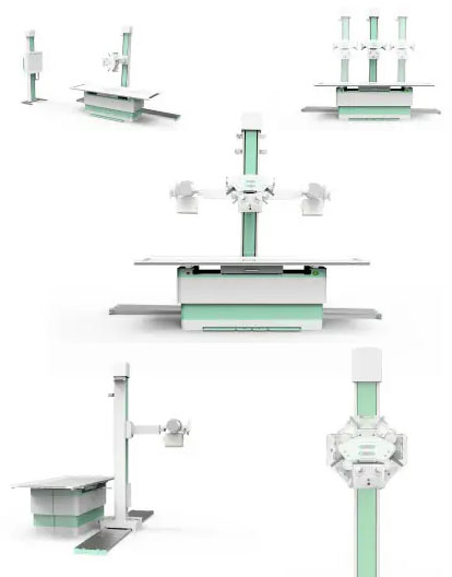
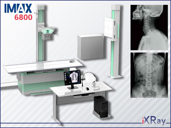
High frequency digital radiography system equipment

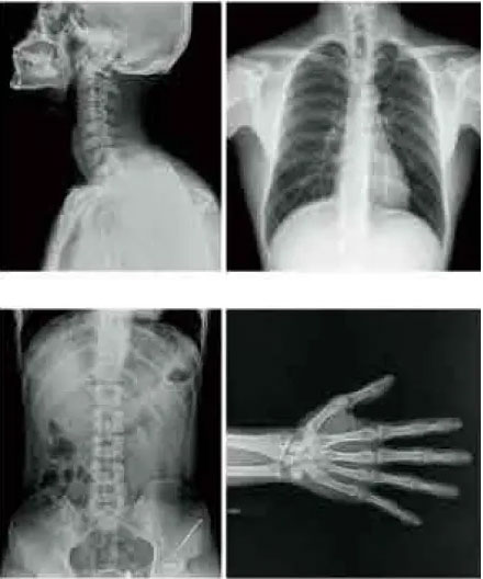
Digital system for different anatomic regions, such as head, chest, abdomen, limbs etc.

Futures & Benefits

Image
Image
Image
Image

HF x-ray generator, 

short time exposure
Image

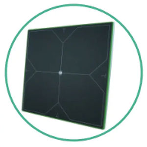
Configuration (1) portable
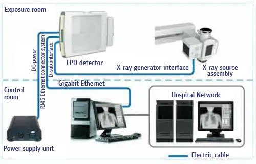
flat panel detector
Image

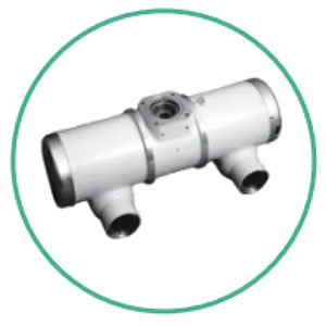
High thermal capacity

of x-ray tube

Image
Image
Image

Convenient digital image network 

Excellent digital imaging processing system for more efficient workflow. 

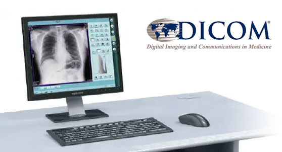
DICOM 3.0 networking interface for seamless integration into your PACS or RlS system

● DigitalStorage: quick and convenient, save the cost comparing with traditional film storage

● lmage process: multi-grade denoise, LlH, negative & positive images, image reverse and so on

● Medical history management: database management, graphicreports, support worklist

● Digital Printer: can be connect with laser printer to print film.

● Digital Network:Support DICOM 3,0 and PACSnetwork in hospital.

● Tissue equalization

● Preset several examination modes to meet different departments use

● ESA (exam specific algorithm)

● Optional: AEC (Automatic exposure control) ensures auto selection of radiographic factors, saves time, eliminates retakes, increases diagnostic capability and lower the radiation dose.

X-ray diagnostic system IMAX 6800
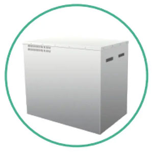
High-frequency X-ray power supply device with a microprocessor control and self-diagnosis system
  • power supply network 380 V, three phases
  • power 80 kW
  • frequency 440 kHz
  • voltage range 40-150kV
  • anode current range 10-900mA
  • mAs range 0.1-1000
  • time range 0.001-10 sec
  • tube protection system against overload
  • 1000 anatomical programs
  • 2-point programming system - kV-mAs
  • 3-point programming system - kV-mA-msec
  • AEC (Automatic Exposure Control)
X-ray table of images
  • the table top is covered with impact-resistant glass
  • the maximum weight of the patient is 220 kg
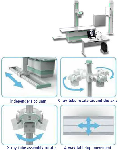
  • floating table top in 4 directions with electromagnetic brakes
  • changing the height of the table by 50 cm
  • the deck size is 2154x860
  • total filtration of 0.5 mmAl
  • sizes of supported cassettes are from 13x18 to 43x43 cm
  • vertical movement of the emitter along the column of 110 cm
  • the rail allows the column to move further than the table, this allows you to take pictures outside the workplaces
  • rotation of the column with the emitter +/-1200
Vertical photo rack
  • fixation of the cassette receiver is carried out with the help of electromagnetic brakes
  • sizes of supported cassettes are from 13x18 to 43x43
  • focal length 1500 cm
X-ray emitter
  • sizes of focal spots 0.6x0.6 mm and 1.2x1.2 mm
  • the maximum voltage on the tube is 150 kV
  • the angle of inclination of the anode is 12 degrees
  • anode rotation speed 9600 revolutions/min
Workstation with a flat panel detector
  • flat panel detector is wireless
  • distribution capacity of 3.5 par.l/mm
  • size 43x43 cm
  • 19-inch monitor (23"’ or 27" option)
  • management of the patient database
  • correction of histograms
  • scaling and cropping of images
  • brightness / contrast
  • negative/positive
  • printing and recording on CD/Flash
  • full support of DICOM 3.0

Information

All images are for demonstration purpose only. You will get the demo images with the QuickStart pack.

Also, all the demo images are collected from Unsplash. If you want to use those, you may need to provide necessary credits. Please visit Unsplash for details.

FacebookLinkedIn