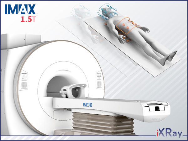
IMAX 1.5T
Magnetic Resonance Imaging System
Futures & Benefits
Focused on excellent performance, Magnetic Resonance Imaging System IMAX 1.5T perfectly meets your needs of quantitative study in MRI practice - with new generation of quantitative analysis tools to fulfill precision medicine and latest applications to broaden your clinical scope.
Also with advanced MUSIC technology, it enables fast image acquisition and multiple exams without repositioning.
See more with "MUSIC" technology and optimized imaging
● The new MUSIC technology advances MR imaging, and Tornado technology reduces motion artifacts.
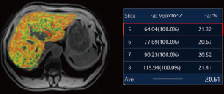
● The PDFF and CQ provide quantitative information for your MRI practice.
Do more with brand-new software platform
● A brand-new iXRay-station is provided for the MR system, users-friendly.
Expect more from our services
● With 12 years of proven user acceptance, international spare parts centres and professional transportation container.
You can expect more from your scanner.
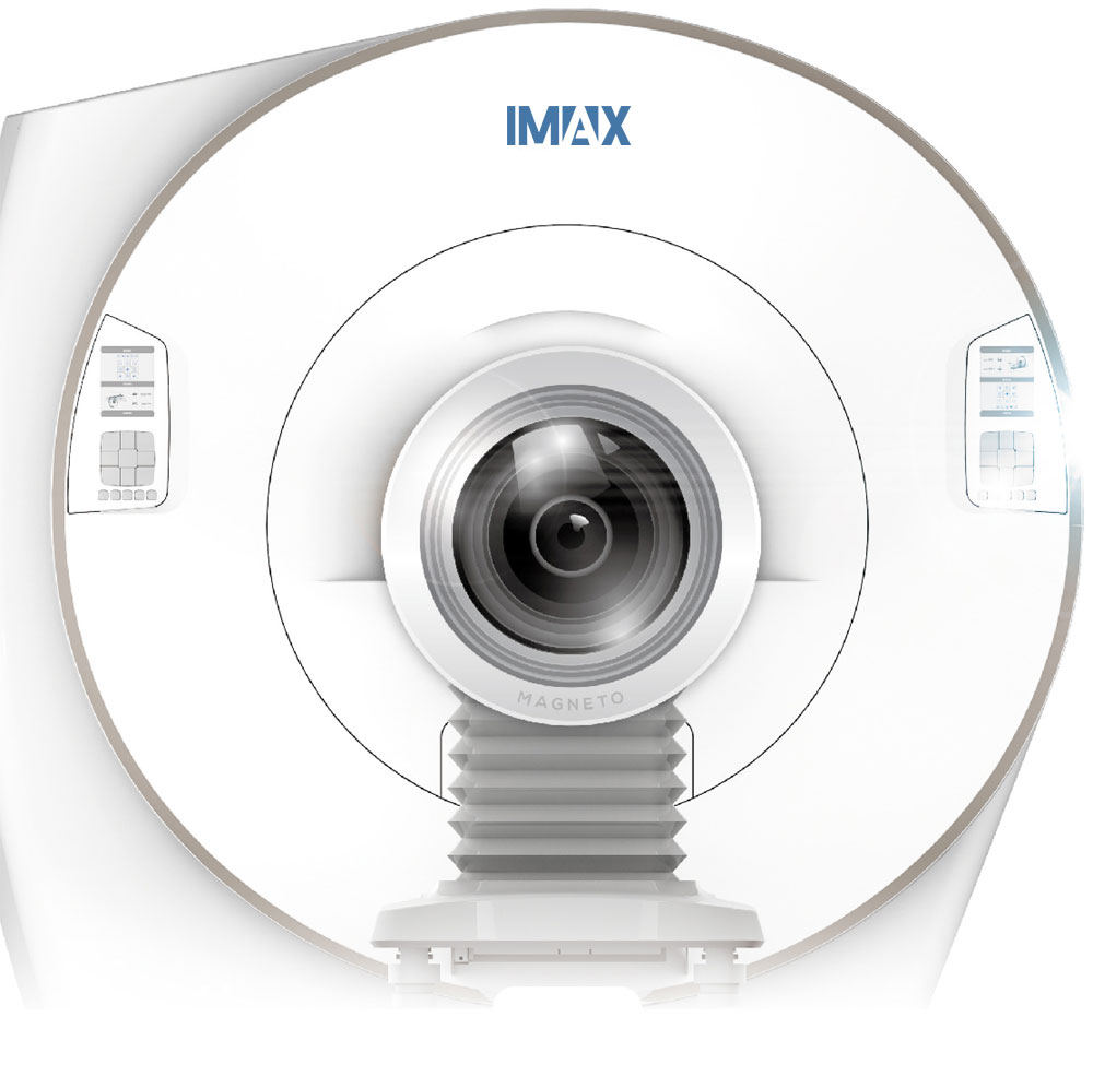
MUSIC
MUSIC (MUiti-Segment Imaging Combination) improves MR imaging with flexibility, precision, and speed. It perfectly integrates a maximum number of 66 channels and provides 16 independent RF channels to be used simultaneously in one single scan and in one FOV. It enhances image quality and acquisition speed to a brand new level. With MUSIC's body coverage, repositioning patients for multiple exams is no longer necessary.
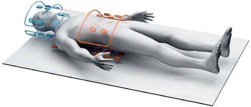
Flexibility
MUSIC is easy to use with more adaptability and versatility. You only need to choose the examination you want without the coils replacement, which improves workflow and increases productivity.
Precision
With excellent and pinpointed precision, MUSIC provides excellent image quality from small lesions to the whole body.
Speed
With MUSIC, the examination set-up is faster and simpler, and acquisition time is shorter. Now patient volume can really skyrocket.
More Imaging
●●● Neuro Imaging ●●●
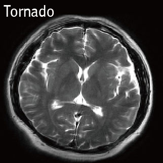
IMAX 1.5T delivers excellent image quality in nervous system. The system supports the complete range of clinical applications. Also many advanced imaging sequences like SWAPP, Tornado can be performed on our system.
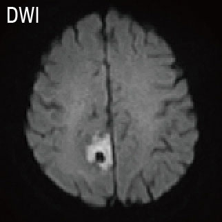
DWI
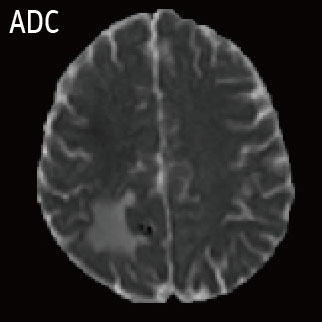
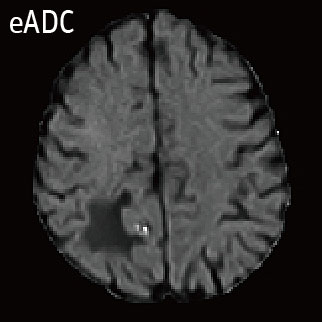
The DWI technique supports multiple b-values and acquired ADC and eADC by post-processing, helps to diagnose super early cerebral ischemia.
SWAPP
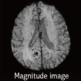
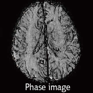
SWAPP is an imaging technique that helps visualize and clearly represent small vessels and microbleeds, as well as large vascular structures, and iron or calcium deposits in the brain. It can generate magnitude image and phase image for diagnosis automatically.
Tornado
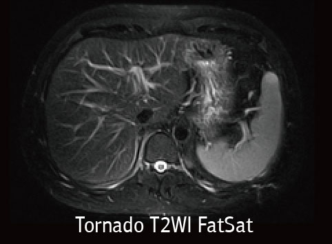
Tornado is designed to reduse the effect of patient voluntary and physiologic motion (respiration, flow, peristalsis) and help visualize the smallest lesion in non-cooperative patients.
●●● Body Imaging ●●●
IMAX 1.5T gets the whole picture with the comprehensive body imaging solutions - a lot of advanced tools designed for your patients.
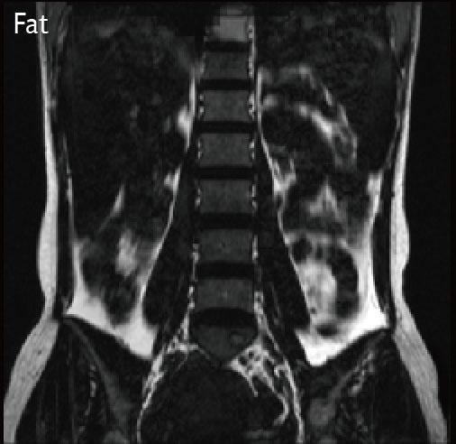
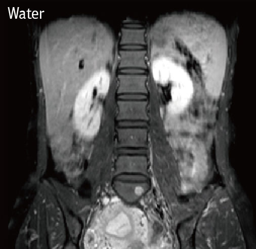
PDFF
PDFF (proton density fat fraction) is a non-invasive imaging method to provide quantitative measurement of hepatic fat content only in 19 seconds.
The methodology in particularly appealing for the pediatric population because of its rapidly and radiation-free imaging techniques.Contrast Enhanced Body imaging
It provides whole-abdominal coverage at hight resolution in short breath-holds, with excelent fat suppresion and resolution.
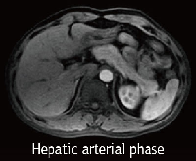
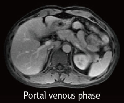
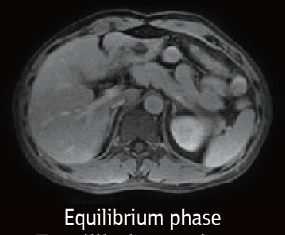
Tornado. Tornado is designed to reduce the effect of patient voluntary and physioligic motion.
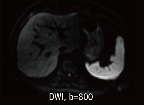
DWI. DWI with b-value 800 helps to diagnose for early liver cancer.
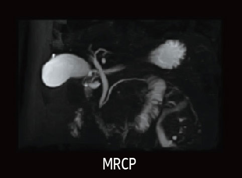
MRCP. High-resolution reliable visualization of the biliary ducts.
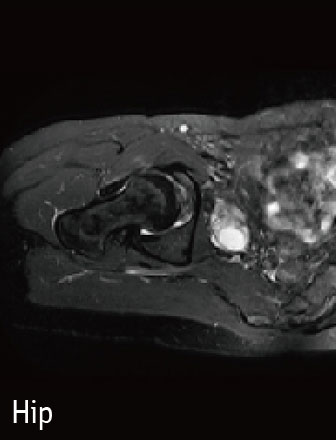
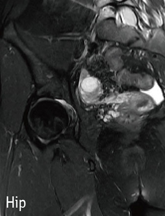
●●● MSK Imaging ●●●
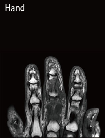
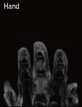
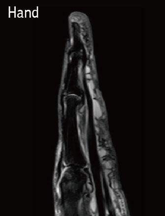
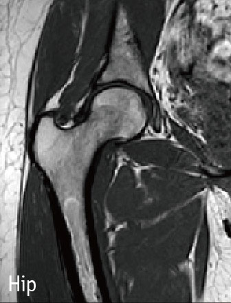
Thanks to a combination of good MSK sequences, advanced RF coils, better computing technologiy and optimazed magnet homogeneity, IMAX 1.5T delivers high resolution MSK images with good acquisition times.
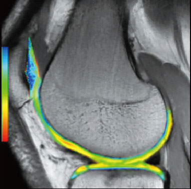
Cartilage Quantification
Cartilage Quantification provides quantitative assessment of cartilage composition to trtack the degradation of tissues in the early stage within joints, which can't be detected by conventional imaging techniques. It allows for noninvasive measurement of collagen content.
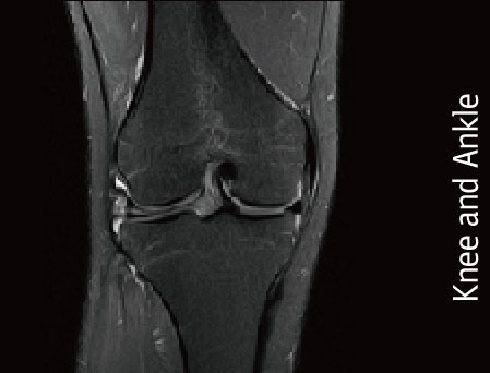
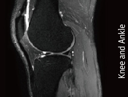
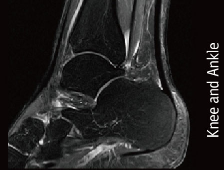
MSK images
This musculoskeletan imaging techniques enables you to image bone, joint and soft tissue with remarcable tissue contrast.
●●● Spine Imaging ●●●
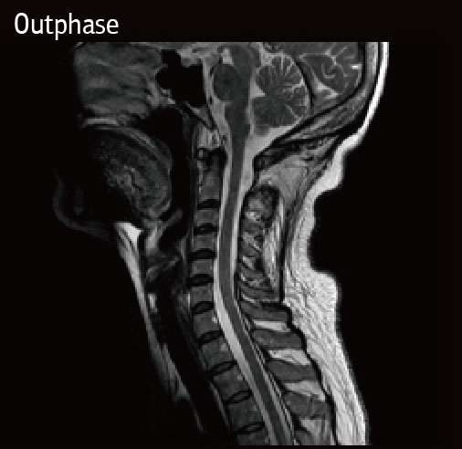
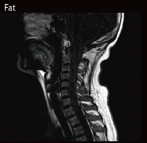
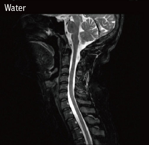
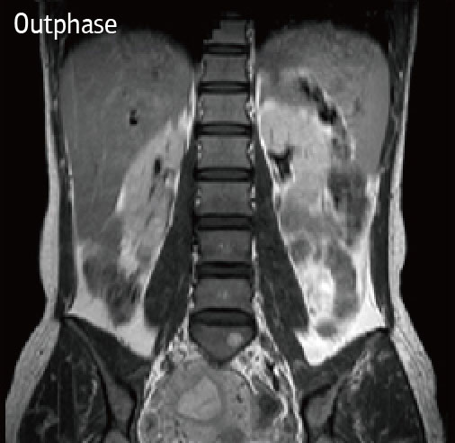
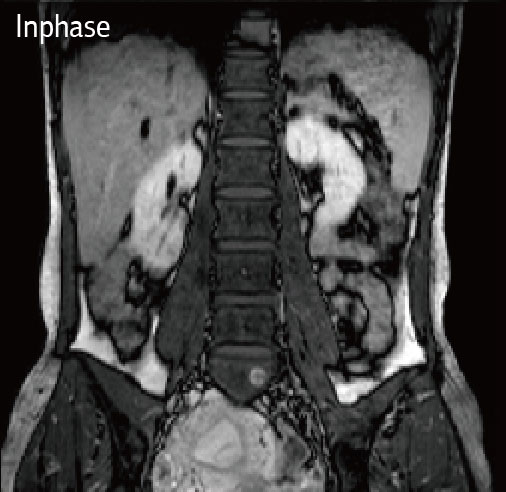
With MUSIC technology, the whole spine imaging can be achieved, which proved useful in the identification of occult vertebral dyspasia and in demonstration of intraspinal and paraspinal neoplasms.
Cervical
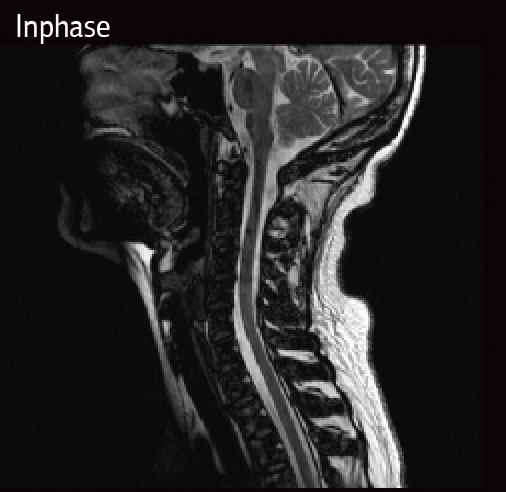
Lumbar
●●● Breast Imaging ●●●
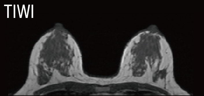
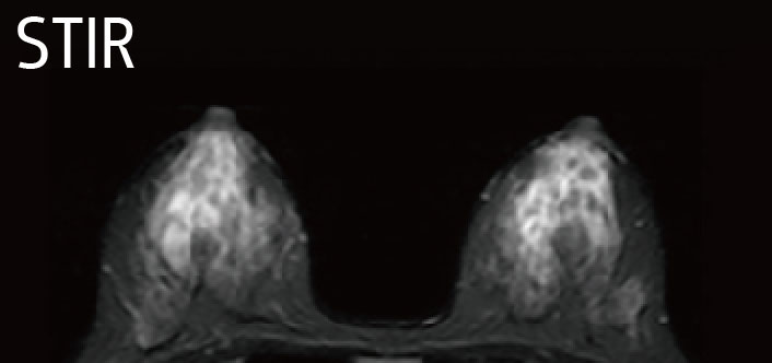
Not all breast MR needs are the same - and neither are all breast MR imaging solutions. With applications and coil designed specifically for breast MR, IMAX 1.5T offers you the best choice.
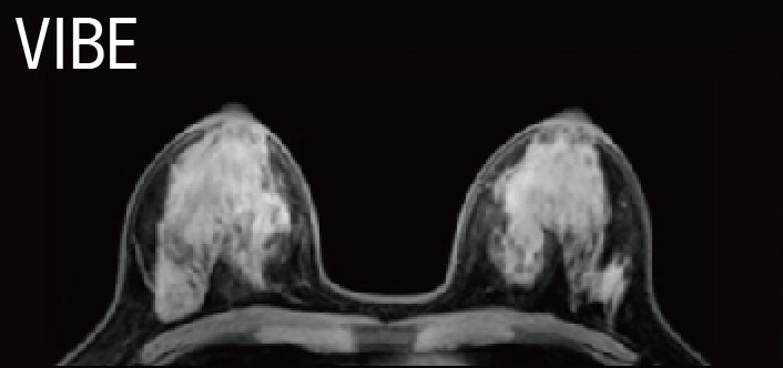
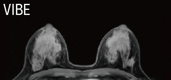
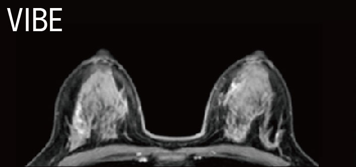
VIBE
VIBE lays foundation of breast MRI with more details and scanning speed. The bilateral shimming ensures uniform bilateral fat saturation. Scan both breasts in one fast exam to help increase diagnostic confidence and patient comfort.
●●● Advanced Imaging ●●●
Offering advanced diagnostic applications is good for patients, who will benefit from the applications of the news tools, and also good for you practice, for it will win a good reputation among customers and patients as the place to solve diagnostic challenges.
rFOV DWI
rFOW DWI can be applied on spine, uterus and prostate, and increases clinical confodence in the diagnosis of numerous common pathologies.
Enhance Inflow IR
Consistent and reiable non-contrast, free-breathing of the arterial and venous vascular, such as the renal and portal vein.
Single Voxel MRS
MRS is ideally suited for measuring therapeutic outcames by obtaining chemical signals from a region of interest.
DCE-MRI
Due to its quick scanning, dynamic enchanced MRI in pituitary can better reflect the changes of its blod supply, improves the detection rate of microadenomas.


