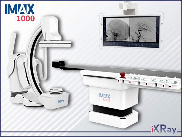
Angiography system IMAX 1000
Futures & Benefits
Digital subtraction angiography system application
● It is suitable for multidisciplinary intervention, such as cardiovascular Surgery; neurosurgery; peripheral vessels; oncology; hepatobiliary surgery; gastroenterology; orthopedic; emergency; radiology; gynaecology; surgery.
● Easily cover different projection parts and meet surgical needs of whole body scanning.
● Compatiable flat panel detector can adopt to different surgeries, such as cardiac intervention, neurointervention.
● 58-inch monitor can display the real-time image and reference image at the same time, and realize 1-4 screen display at the same time.
● Realize one-key operation to the specific position.
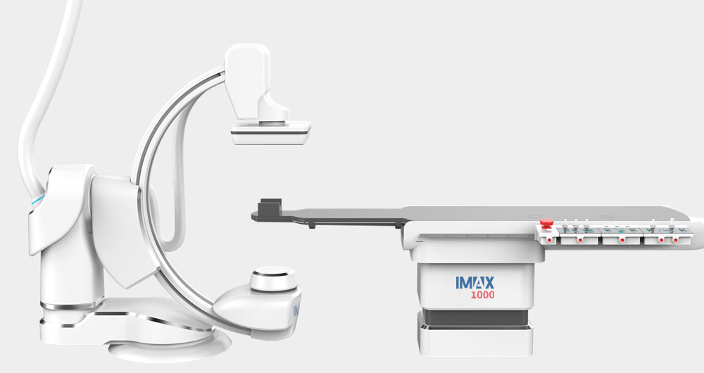
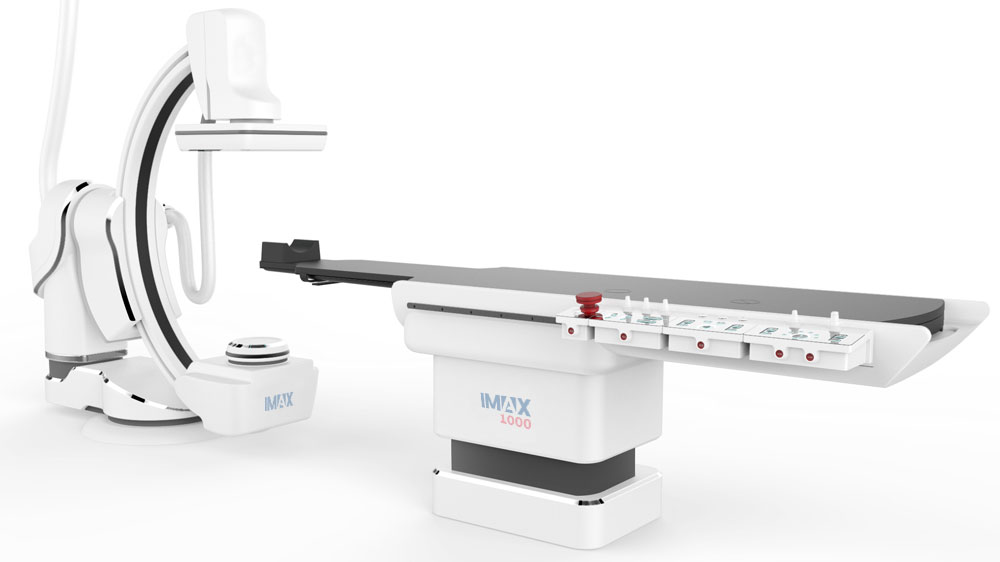
Artificial Intelligence
● Humanized mechanical design scheme
● Intelligent human-computer interaction mode
● Memory private custom service
IMAX 1000's rack design mimics a whale shape, and when combined with a rotating DSA function for multi-angle acquisition and an intelligent algorithm for 3D model reconstruction, doctors can easily find hidden lesions. The entire 16bit + 2K digital image chain, as well as the 5A intelligent algorithm, can easily complete the operation in low-dose mode and obtain high- definition images to help optimise, control and monitor the intelligent dose.
IMAX 1000 is made from...
● High-resolution image engine
● Ultrasmart gantry movement
● "We Dose" micro-dose protection
● One-click operation
● Multi-customised clinical applications
● Liquid metal bearing tube with dual cooling system
● Smart floating table in all directions
● 58-inch HR 4k display.
Angiography system IMAX 1000 provides clear imaging for a variety of clinical procedures and good visibility for patients of all sizes at low X-ray dose levels. To enhance user experience and decision-making, users can control the app via a central touchscreen and large screen solution next to the examination table, allowing doctors to evaluate and make decisions in the sterile field while saving time and avoiding delays. The friendly man- machine bedside operation panel and intelligent microcomputer simplify and standardise system setting and setup from routine to complex surgery, and provide clinical tools to perform surgery.
An integrated portfolio of technologies and services supports a wide range of interventional procedures and provides a combined operating room solution in an ideal care environment for performing open and minimally invasive surgical procedures. Other features include real-time image guidance, including stent fine imaging, CT-like functions and navigation guidance solutions, such as aortic valve replacement navigation. All functions can be integrated into the system to support clinical workflow.
Angiography system IMAX 1000’s rack design mimics a whale shape, and when combined with a rotating DSA function for multi-angle acquisition and an intelligent algorithm for 3D model reconstruction, doctors can easily find hidden lesions. The entire 16bit + 2K digital image chain, as well as the 5A intelligent algorithm, can easily complete the operation in low-dose mode and obtain high- definition images to help optimise, control and monitor the intelligent dose.
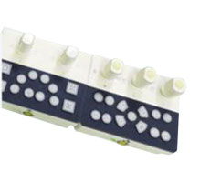
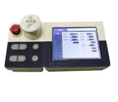
Table-side microcomputer unit "Smart Box"
● Gantry position automatic control
● Image acquisition control
● Image Postprocessing control
Ultrasmart gantry movement
● 7 axes synchronized movement
● Customized one-click position svitching
● Suitable for hybrid operations
● 4 level anti-collision protection

Multifunctional footswitch
● Easy to use
● Easy to desinfection
● Easy to put back
● Multifunctional

● Auto-contrast ● Auto-brightness ● Auto-sharpness ● Auto-dose ● Auto-balance

"Cloud" services
● Image cloud ● Device cloud ● Data cloud
| IMAX 1000 angiographic system | |
| General characteristics |
|
| X-ray table of images: |
|
| C-arc |
|
| X-ray emitter |
|
| Workstation with a flat panel detector |
|
| Display system |
|
| Software for data management |
|
| Software for image storage and transfer |
|
| Basic package of 3D programs | |
| A specialized software package for neurosurgery |


